Quantitative detection of fibronectin in porcine small intestinal submucosa extracellular matrix
作者:
陈毅,夏磊磊,张扬,高建萍,李赛娜,雷雄心,李勇超,张贵锋,赵博
中文摘要:
采用分批次酶解和高效液相色谱与质谱分析联用技术(HPLC-MS)对脱细胞猪小肠黏膜下层(p-SIS)基质材料中纤维连接蛋白(FN)进行了定量检测。分批次酶解消除了静态酶解时间长、酶解效率低的不足,将酶解时间从96 h缩短至6 h,降解率可以达到95%以上。采用胰蛋白酶特异性酶解得到猪源FN的一个特征多肽为SSPVVIDASTAIDAPSNLR,HPLC-MS对这一特征肽段定量检测结果表明,VIDASISTM和Biodesign®两种脱细胞材料中FN的含量分别为0.43%和0.17%。方法学实验证明该方法精确度较高(RSD为1.31%)。结果表明基于质谱的分析技术可有效检测脱细胞基质中FN含量。
英文摘要:
Quantitative analysis of fibronectin in porcine small intestinal submucosa(p-SIS) acellular matrix was investigated using batch enzymatic digest and high performance liquid chromatography mass spectrometry (HPLC-MS). The results indicated that batch enzymatic digest could improve the hydrolysis efficiency compared to static enzymatic hydrolysis. The hydrolysis time decreased to 6 hours from 96 hours, and the enzymatic hydrolysis rate amounted to 95%. Marker peptide, SSPVVIDASTAIDAPSNLR, was detected from the tryptic digest of FN. Based on the peak areas of marker peptide from FN standards with different concentration, the FN content in VIDASISTM was determined as 0.43%. Amount of FN in Biodesign® was determined as 0.17%. The method precision (RSD, 1.31%) and the deviation were carried out in the methodology experiments. The results indicate the established quantitative method is effective for detecting FN content in acellular matrix.
关键词:
猪小肠黏膜下层;;脱细胞基质;纤维连接蛋白;特征肽段;液质联用技术
Keywords:
porcine small intestinal submucosa; extracellular matrix; fibronectin; marker peptide; LC-MS
生物学杂志,第35卷第2期,2018年4月

对一个家庭来说,最大的喜事之一,莫过于添...
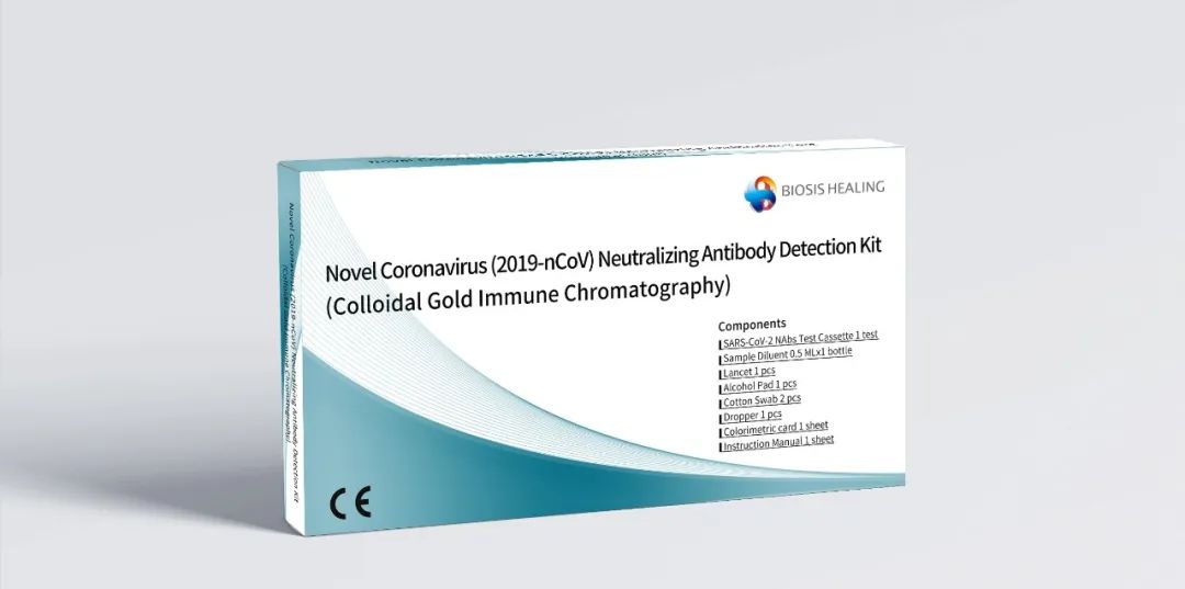
截止目前,全球已有十余款新冠疫苗在多个国...
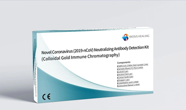
产品名称:新型冠状病毒(2019-nC...

正文...

南德 TÜV CE证书编号:No.G2...

南德 TÜV CE证书编号:No.G2...

南德 TÜV CE证书编号:No. G...

南德 TÜV CE证书编号:No.G2...
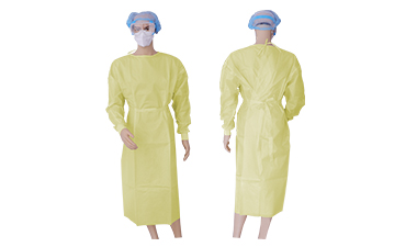
南德 TÜV CE证书编号:No. G...

南德 TÜV CE证书编号:No. G...
版权所有北京博辉瑞进生物科技有限公司 京ICP备16026579号 (京)-非经营性-2017-0030  京公网安备 11011502006176号 (京)网药械信息备字(2022)第00096号